
| Added | Thu, 05/01/2017 |
| Источники | О. А. Антонова, Возрастная анатомия и физиология, Изд.: Высшее образование, 2006 г.
Лысова Н. Ф. Возрастная анатомия, физиология и школьная гигиена. Учеб. пособие / Н. Ф. Лысова, Р. И. Айзман, Я. Л. Завьялова, В.
Погодина А.Б., Газимов А.Х., Основы геронтологии и гериатрии. Учеб. Пособие, Ростов-на-Дону, Изд. Феникс, 2007 – 253 с.
|
People cannot see in complete darkness. In order that the person saw the object, it is necessary that the light reflected from the object onto the retina of the eye. The light sources can be natural (fire, Sun) and artificial (various lamps). But what is the light?
According to modern scientific ideas, light is a electromagnetic wave of a certain (rather high) frequency range. This theory originates from Huygens as confirmed by many experiments (in particular, experience T. Jung). In the nature of light to fully manifest karpuskulyarno wave-particle duality, which largely determines its properties during the propagation of light behaves like a wave in the emission or absorption – as a particle (photon). Thus, light effects occurring during the propagation of light (interference, diffraction etc.) are described by Maxwell's equations, and the effects that occur when the absorption and radiation (photoelectric effect, Compton effect) – the equations of quantum field theory.
Simplistically, the human eye is a radio receiver capable of receiving electromagnetic waves of a certain (optical) frequency range. The primary sources of these waves are body, their radiant (sun, lamps, etc.), the secondary – body, reflecting the wave of primary sources. The light from the sources enters the eye and makes them visible to man. Thus, if the body is transparent to waves in the visible frequency range (air, water, glass, etc.), it cannot be registered by the eye. With the eyes, like any other radio receiver "tuned" to a specific range of radio frequencies (in the case of the eye is in the range of from 400 to 790 terahertz), and does not take waves having a higher (ultraviolet) or low (infrared) frequency. This "setting" is evident throughout the structure of the eye from the lens and vitreous, transparent in this frequency range, and ending with the amount of photoreceptors, which in this analogy is similar to radios and antennas have dimensions that ensure the most efficient reception of radio waves of this range.
All this together defines the range of frequencies in which to see people. He called the range of visible radiation.
Visible radiation — electromagnetic waves perceived by the human eye, which is a part of the spectrum with wavelengths from approximately 380 (violet) to 740 nm (red). These waves occupy the frequency range from 400 to 790 terahertz. Electromagnetic radiation with such frequencies, also called visible light or simply light (in the narrow sense of the word). The highest sensitivity to light of human eyes is in the region of 555 nm (540 THz) in the green part of the spectrum.
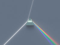
White light separated by a prism into the colors of the spectrum [4]
The decomposition of a ray of white color in the prism spectrum is formed in which the radiation of different wavelengths are refracted at different angles. The colors included in the spectrum, i.e. those colors that can be obtained with the light waves of the same length (or very narrow band) are called the spectral colors. The basic spectral colors (with own name), as well as the radiation characteristics of these colors are presented in the table:

The spectrum does not contain all the colors that distinguish the human brain and they are formed from mixing other colors.[4]
What a person sees
Through sight we receive 90% of information about the world, so the eye is one of the most important senses.
The eye is a complex optical instrument. Its main task is to "transfer" the right image to the optic nerve.

The structure of the human eye
The cornea is transparent shell covering the front part of the eye. It lacks blood vessels, it has a large refractive power. Included in the optical system of the eye. The cornea is bordered with an opaque outer shell of the eye — sclera.
Anterior chamber is the space between the cornea and iris. It is filled with intraocular fluid.
Iris looks like a circle with a hole inside (pupil). The iris consists of muscles, while reducing and relaxing the size of the pupil change. It is part of the choroid of the eye. Iris is responsible for eye color (if it is blue then it contains few pigment cells, if brown — a lot). Performs the same function as the aperture in the camera, adjusting svetotochek.
Pupil — hole in iris. Its size usually depends on the level of illumination. The more light the smaller the pupil.
The lens is a "natural lens" of the eye. It is transparent, elastic — can change its shape almost instantly "directing focus", whereby one can see well both near and far. Within the capsule is retained ciliary zonule. The lens, like the cornea, is included in the optical system of the eye. The transparency of the lens of the human eye is excellent - skipped most of the light with wavelengths between 450 and 1400 nm. Light with a wavelength of nm выше720 not perceived. The lens of the human eye is almost colorless at birth, but turn yellowish with age. It protects the retina from ultraviolet rays.
Vitreous body — a transparent gel-like substance located in the posterior part of the eye. The vitreous body maintains the shape of the eyeball and is involved in intraocular metabolism. Included in the optical system of the eye.
Retina consists of photoreceptors (they are sensitive to light) and nerve cells. Cell receptors located in the retina, are divided into two types: the rods and cones. In those cells that produce the enzyme rhodopsin converts light energy (photons) into electrical energy of the nervous tissue, i.e. photochemical reaction.
Sclera — opaque outer shell of the eyeball, passing in front of the eyeball in a transparent cornea. Attached to the sclera 6 of the eye muscles. It is a small number of nerve endings and blood vessels.
Choroid — lines the posterior sclera, it adjoins the retina, with which it is closely connected. Choroid is responsible for blood supply of intraocular structures. In diseases of the retina very often involved in the pathological process. In the choroid has no nerve endings, so when it is the disease not have pain, usually signaling any failures.
Optic nerve — with the help of the optic nerve signals from the nerve endings are transmitted to the brain.[6]
Man is not born with an already-developed organ of vision: in the first months of life is the formation of the brain and vision, and by about 9 months they are able almost immediately to process the incoming visual information. In order to see, light is needed. [3]
The light sensitivity of the human eye
The eye's ability to perceive light and recognise different levels of brightness is called light perception, and the ability to adapt to different lighting brightness adaptation of the eye; light sensitivity is evaluated by the threshold value of the light stimulus.
A person with good vision can see at night the light from the candle at a distance of several kilometers. The maximum light sensitivity is reached after a sufficiently long dark adaptation. It is determined, under the action of a luminous flux in a solid angle 50° at a wavelength of 500 nm (maximum sensitivity of the eye). Under these conditions, the threshold energy of the light is about 10-9 erg/s, which is equivalent to the stream of several photons of optical range per second through the pupil.
Contribution of the pupil to adjust the sensitivity of the eye is extremely low. The full range of light to dark that our eye can perceive, from 10-6 CD•m2 for the eye is fully dark-adapted, to 106 CD•m2 for the eyes, completely adapted to the world Mechanism of this broad range of sensitivity lies in the decomposition and reconstruction of the photosensitive pigments in the photoreceptors of the retina — cones and rods.
In the human eye contains two types of photosensitive cells (receptors): highly sensitive sticks responsible for twilight (scotopic) vision, and less sensitive cones are responsible for color vision.
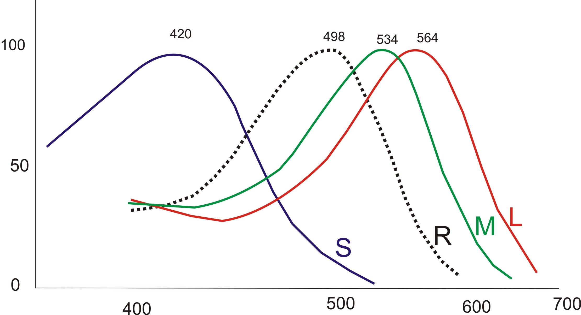
Normalized graphs of the light sensitivity of cones of the human eye S, M, L. the Dashed line shows the twilight, "black and white" the sensitivity of the sticks.
In the human retina there are three kinds of cones, the peaks of sensitivity which are in red, green and blue parts of the spectrum. The distribution of types of cones in the retina unevenly: the "blue" cones are closer to the periphery, while the "red" and "green" are distributed randomly. Compliance types of cones the three primary colors enables recognition of thousands of colors and shades. Curves spectral sensitivity of the three types of cones overlap, which contributes to the phenomenon of metamerism. Very strong light excites all 3 types of receptors, and therefore is perceived as radiation blindingly white.

Uniform irritation all three elements, corresponding to the weighted average daylight also causes the sensation of white.
For color vision person responsible, genes encoding light-sensitive proteins, the opsins. According to supporters of the three-component theory, the presence of three different proteins that respond to different wavelengths, is sufficient for color perception.
Most mammals these genes only two, so they have black and white vision.
Sensitive to red light opsin is encoded in human OPN1LW gene.
Other human opsins encode genes OPN1MW, and OPN1SW OPN1MW2, the first two of them encode proteins that are sensitive to light of middle wavelengths, and the third is responsible for the opsin sensitive to the shortwave part of the spectrum.
The field of view
Field of view — space, simultaneously perceived by the eye with a rigid eye and a fixed position of the head. It has certain boundaries, corresponding to the optical transition of the active part of the retina in optical blind.
The field of view is artificially limited to protruding parts of the face — nose, the upper edge of the orbit. In addition, it depends on the position of the eyeball within the eye socket. [8] in addition, in each eye of a healthy person there is a region of the retina, not sensitive to light, called the blind spot. Nerve fibers from the receptors to the blind spot are on top of the retina and are collected in the optic nerve, which passes through the retina on the other side. Thus, in this place no light receptors.[9]
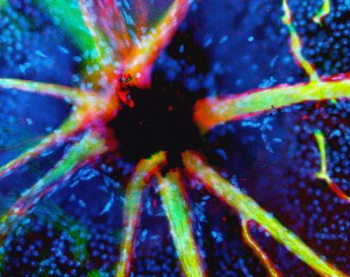
This confocal micrograph of optic nerve shown in black, cells lining the blood vessels is red, and the contents of the vessels green. Cells in the retina display blue spots. [10]
Blind spots in the two eyes are in different places (symmetrically). This fact, as well as the fact that the brain adjusts the perceived image, explain why during normal use of both eyes they are invisible.
To see in his blind spot, close the right eye and left eye look at the right cross, which is circled. Keep your face and the monitor vertically. Without taking his glance from the right cross, zoom (or Attaleia) a person from the monitor and at the same time watch out for the left cross (without taking her gaze on him). At some point it will disappear.
This way you can also estimate the angular size of the blind spot.

The reception for the detection of the blind spot [9]
There are also departments of the paracentral visual field. Depending on participation in the vision one or both eyes, and can distinguish between monocular and binocular field of view. In clinical practice usually investigate the monocular field of view. [8]
Binocular and Stereoscopic vision
Visual analyzer of the person in normal conditions provides binocular vision, i.e. vision with two eyes with a single visual perception. Reflex the main mechanism of binocular vision is a reflex of merge image fusion reflex (fusion) created by simultaneous stimulation of varies functional nervous elements of the retina of both eyes. The result is a physiological double vision of objects located closer to or further fixed points (binocular focus). Physiological double vision (focus) helps to estimate the distance of the object from the eye and creates a sense of relief or stereoscopic, vision.
When vision one eye the depth perception (the relief distance) is mainly due to the secondary auxiliary signs of distance (the apparent size of the subject, linear and aerial perspective, occlusion of some objects by others, the accommodation of the eye, etc..). [1]
The conducting tracts of visual analyzer
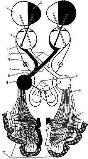
1 — Left half of the visual field, 2 — Right half of the visual field, 3 — Eyes, 4 — Retina, 5 — Optic nerve, 6 — Oculomotor nerve 7 — the Chiasm, The 8 Optic tract, 9 — Lateral geniculate body, 10 — Top bumps of the corpora quadrigemina, 11 — non-specific visual pathway, 12 — the Visual cortex of the brain.[2]
Man sees not with the eyes but through the eyes, where information is transmitted through the optic nerve, optic chiasm, optic tracts in certain areas of the occipital lobes of the cerebral cortex, where there is the picture of the external world that we see. All these organs make up our visual analyzer or visual system.[5]
Vision changes with age
The elements of the retina begin to form at 6-10 week of fetal development, the final morphological maturation occurs by the age of 10-12. In the development process of the body changing the color perception of the child. The newborn in the retina are sticks that provide black and white vision. The number of small cones and they are still not Mature. Color recognition at an early age depends on luminance, not from the spectral characteristics of the color. As maturation of the cones children first distinguish yellow, then green and then red (with the 3 months were able to develop conditioned reflexes for these colors). Full cones start functioning by the end of 3 years of life. At school age distinctive color sensitivity of the eye increases. Maximum development of the sensation of colour reaches the age of 30 and then gradually decreases.
Newborn diameter of the eyeball is 16 mm and its weight is 3.0 g. the Growth of the eyeball continues after birth. Most intensively it grows the first 5 years of life, less intensively, up to 9-12 years. Newborns form of the eyeball is more globular than in adults, resulting in 90% of cases they observed sighted refraction.
The pupil in newborns narrow. Due to the prevalence of tonus of sympathetic nerves innervating the muscles of the iris, is 6-8 years the pupils become wide, which increases the risk of sunburn of the retina. In 8-10 years the pupil narrows. In 12-13 years, the speed and intensity of the pupillary response to light becomes the same as that of an adult.
In infants and children of preschool age, the crystalline lens more convex and more elastic than the adult, its refractive power higher. This allows the child to clearly see an object at a shorter distance from the eyes than an adult. And if the baby it is transparent and colorless, in the adult, the lens has a light yellowish tint, the intensity of which with age may increase. It does not affect visual acuity but may affect the perception of blue and purple flowers.
Sensory and motor functions of vision developed simultaneously. In the first days after birth eye movement out of sync, with immobility of one eye to observe the other. The ability to fix the gaze of the subject is formed at the age from 5 days to 3-5 months.
Reaction to the shape of the object is observed in 5-month-old baby. In preschool children the first reaction causes the shape of the object, then its size, and especially color.
Visual acuity increases with age, and improved stereoscopic vision. Stereoscopic vision for 17-22 years reaches its optimal level and with 6 years in girls, the sharpness of stereoscopic vision is higher than that of boys. The field of view is growing vigorously. To 7 years it amounts to approximately 80 % of the size of the field of view of an adult.[11,12]
After 40 years there is a decline in the level of peripheral vision, then there is the narrowing of the field of view and the deterioration in the lateral view.
After about 50 years decreases production of the tear fluid, so eyes moistened worse than in younger age. Excessive dryness may result in redness of the eyes, cramps, watery eyes due to wind or bright light. It may not depend on the usual factors (frequent eye strain or air pollution).
With age, the human eye begins to perceive the environment more dim, lower contrast and brightness. Can also deteriorate the ability to recognize shades of color, especially close in color. This is directly related to a reduction in the number of cells in the retina that perceive color, contrast, brightness. [14,15]
Some age-related blurred vision due to presbyopia, which is manifested by vagueness, blurring of the images when you try to examine objects close to the eye. The ability to focus vision on small objects requires the accommodation of about 20 dioptres (focusing on an object 50 mm from the observer), to 10 dioptres at age 25 (100 mm), and levels of 0.5 to 1 dioptre at age 60 (ability to focus on the subject of 1-2 metres). It is believed that this is due to the weakening of the muscles that govern the pupil, thus also deteriorating the reaction of pupils on entering the eye the light beam. [13] Therefore, having difficulty with reading in dim light and increases during adaptation when changes in light conditions.
Also with age begins to occur faster eye fatigue and even headaches.
Color perception
The psychology of color perception — ability to perceive, identify and name the colors.
The sensation of color depends on the complex physiological, psychological and cultural-social factors. The original study of color perception was conducted in the framework of color; later, the issue is connected ethnographers, sociologists, and psychologists.
Visual receptors are considered "part of the brain rendered on the surface of the body." Unconscious processing and correction of visual perception provides a "correct" view, and it is the cause of "errors" in the evaluation of color in certain conditions. So, the elimination of "background" exposure of the eye (for example, when looking at distant objects through a narrow tube) significantly changes the perception of the colors of these items.
Simultaneous viewing of the same niesamowity of objects or light sources by several observers with normal colour vision, under the same conditions of viewing, allows to establish an unambiguous correspondence between the spectral composition of the compared radiations and caused their color sensations. Based on this color measurement (colorimetry). This correspondence uniquely, but not mutually clear: the same color sensation can cause the flow of radiation of different spectral composition (metamerism).
Definition of color as a physical quantity, there are many. But even in the best of them with a colorimetric point of view is often omitted mention of the fact that this (not mutual) uniqueness is achieved only in standardized conditions of observation, lighting, etc., is not considered a change in the perception of color when you change the intensity of radiation of the same spectral composition (the phenomenon Bezold — Brücke), is not taken into account the so-called color adaptation of the eyes, etc. So the variety of color sensations that arise in actual lighting conditions, variations of angular size to compare the color of elements, their fixation on different parts of the retina, different physiological States of the observer, etc. always richer colorimetric color diversity.
For example, in colorimetry, it equally identifies some of the colors (such as orange or yellow), which in everyday life are perceived (depending on the lightness) as a brown "chestnut", brown, "chocolate", "olive", etc. In one of the best attempts to define the concept of Color, owned by Erwin schrödinger, the difficulties are removed by a simple absence of instructions to the dependence of color sensations from the numerous specific conditions of observation. According to schrödinger, Color is a property of spectral composition of radiations, the General to all radiations, visually indistinguishable to humans. [6]
Due to the nature of the eye, light, evocative of the same color (e.g. white), that is, the same degree of stimulation of the three visual receptors, can have different spectral composition. People in most cases do not notice this effect, as if "conjecturing" color. This is because although different color temperature lighting can coincide, the reflected spectra of the same pigment of natural and artificial light can differ and cause different color feeling.
Human eyes perceive many different shades, but there are "forbidden" colors, not available to him. As example, the color plays and yellow and blue tones at the same time. This is because the perception of color in the human eye, like many other things in our body, built on the principle of opponentsthe. The retina is of particular neurons-opponents: some of them aktiviziruyutsya when we see red, and they are suppressed in green. The same thing happens with a pair of yellow-blue. Thus, the color pairs red-green and blue-yellow have the opposite effect on the same neurons. When the source emits both colors of the pair, their impact on neuron kompensiruet, and people can't see none of these colors. Moreover, people are not only able to see these colors in normal circumstances, but also to present them.
To see these colors only in the framework of a scientific experiment. For example, scientists Hewitt crane and Thomas Piantanida from Stanford Institute in California have created a special visual models that alternated stripes "arguing" shades, quickly replacing each other. These images, recorded by a special device at eye level of the person, were shown dozens of volunteers. After the experiment, the people argued that at some point the border between the shades disappeared, merging in one color, with which previously they never had to face.
The differences of humans and animals. Metamerism in photography
Human vision is tractimeline analyzer, that is, the spectral characteristics of the color expressed by only three values. If we compare the fluxes of different spectral composition are produced on cones of the same action, colors are perceived as identical.
In the animal world there are four and even patestealse color analyzers, therefore, the color perceived by man are the same animal can appear different. In particular, birds of prey can see evidence of rodents on the trails and burrows solely due to the ultraviolet luminescence of the components of their urine.
A similar situation exists with the systems of registration of images, both digital and analog. Although most of them are tractimeline (three layers of film, three types of matrix cells of a digital camera or scanner), metamerism metamerism is different from human vision. Therefore, the color perceived by the eye as equal in the picture may be different, and Vice versa. [7]
Translated by «Yandex.Translator»
Log in or register to post comments